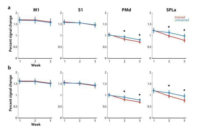
Author response image 3. Elicited activation, measured as percent signal change over resting baseline across weeks for trained (red) and untrained (blue) sequences.

Regions: M1 (primary motor cortex), S1 (primary somatosensory cortex), PMd (dorsal premotor cortex), SPLa (superior parietal lobule – anterior part). a) Estimated elicited activation based on preprocessing, reported in manuscript. b) Estimated elicited activation after removing error trials from a general linear model estimation.
