. 2020 Apr 27;9:e55965. doi: 10.7554/eLife.55965

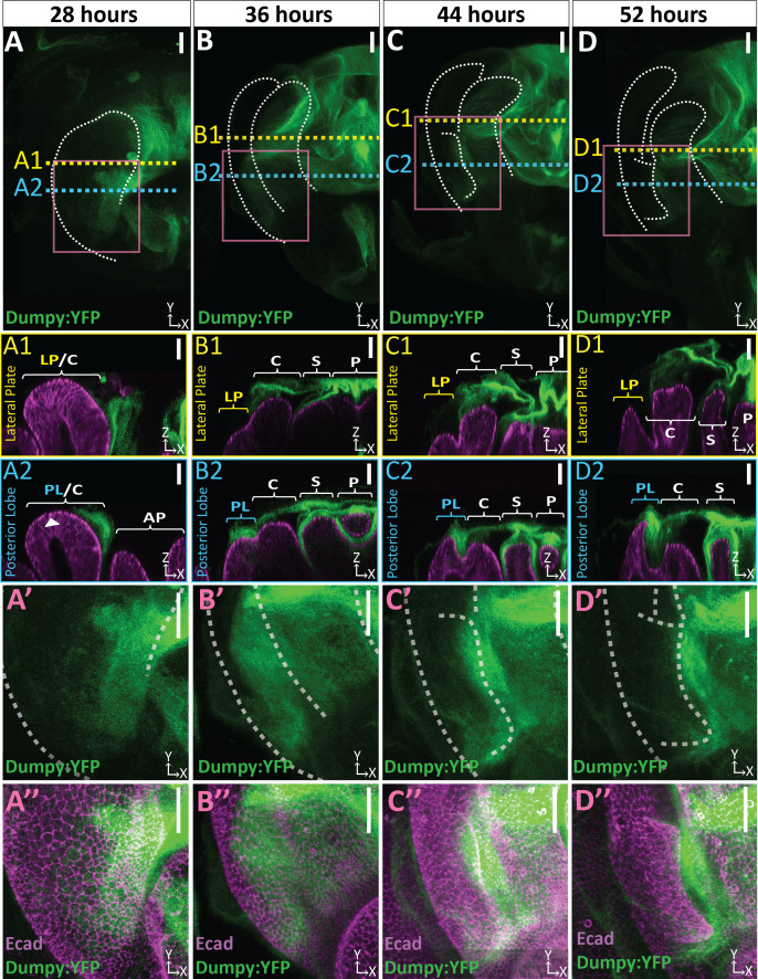
Figure 4. Dumpy deposition is correlated with posterior lobe development.

(A–D) Maximum projection and (A’–B’’) respective zoom, indicated with pink box, labeled with Dumpy:YFP (green) and E-cadherin (Ecad; magenta) for each time point. Location of respective cross sections indicated in yellow for lateral plate (A1–D1) and blue for posterior lobe (A2–D2). Arrowhead in (A2) indicates future posterior lobe cells. Cross-sections are oriented with apical side at the top and basal side at the bottom. Relevant structures labeled: Posterior lobe (PL), lateral plate (LP), clasper (C), sheath (S), and phallus (P). Scale bar, 20 μm. n ≥ 4 per experiment. Images were independently brightened to show relevant structures.

Figure 4.

Figure 4—figure supplement 1. Limited basal ECM present during posterior lobe morphogenesis.

Figure 4—figure supplement 1.

Basal ECM markers Collagen IV (Viking:GFP; green)(A and C) and Perlecan (Perlecan:GFP; green) (B and D) in L3 larval genital disc (A and B) and 44 hr APF genitalia (C and D). Apical membrane labeled with E-cadherin (Ecad; cyan) and lateral membrane labeled with Fasciclin III (Fas3; magenta). Image settings were the same for each marker between larval and pupal samples. Sporadic dots observed are fat bodies (white arrows in cross section), which fill the basal lumen of the pupal genital epithelium. Location of respective cross sections indicated in white. Cross-sections for larval samples are oriented basal sides out, as the disc has not yet everted. Pupal samples are oriented with apical side at the top and basal side at the bottom. Higher amounts of basal ECM are observed in larvae compared to 44 hr APF genital samples. Relevant structures labeled: Posterior lobe (PL) and clasper (C). Scale bar, 20 μm.
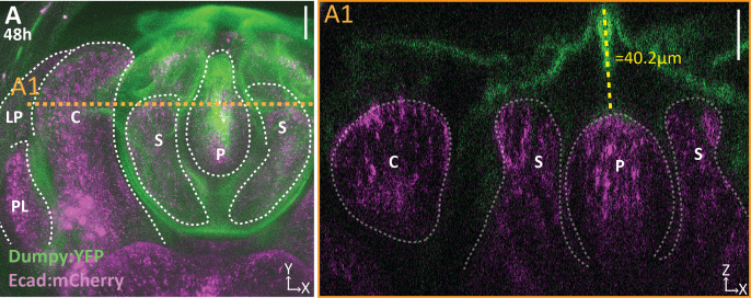
Figure 4—figure supplement 2. Dumpy extends above the apical surface of the phallus.

Figure 4—figure supplement 2.

(A) Projection of Dumpy:YFP (green) and E-cadherin:mCherry (Ecad:mCherry; magenta) imaged live at 48 hr APF. Location of respective cross sections indicated in orange. (A1) Cross section showing extent of Dumpy:YFP observed above the surface of the genitalia. Relevant structures labeled: Posterior lobe (PL), lateral plate (LP), clasper (C), sheath (S), and phallus (P). Scale bar, 20 μm. n = 3.
Figure 4—figure supplement 3. A bundle of Dumpy connects the genitalia to the pupal cuticle membrane that encases the developing pupa.

Figure 4—figure supplement 3.

(A–B) Live imaging of Dumpy:YFP (green) and Ecadherin:mCherry (Ecad:mCherry; magenta) at respective time points. Location of respective cross sections indicated in orange. (A1–B1) Cross-sections are max projection of a 4.94 μm (A1) and 1.73 μm (B1) thick section to show the full bundle (arrow) and its connection to the cuticle (arrowhead) and anal plate. All cross-sections are oriented with apical side at the top and basal side at the bottom. Relevant structures labeled: Posterior lobe (PL), lateral plate (LP), clasper (C), sheath (S), phallus (P), and anal plate (AP). Scale bar, 20 μm. n = 1 per each time point.
Figure 4—figure supplement 4. Weak aECM connections extend to the lateral plate.

Figure 4—figure supplement 4.

(A–D) Brightened images of respective cross sections from Figure 2 of lateral plate (A1–D1) in yellow and posterior lobe in blue (A2–D2). Cross-sections are oriented with apical side at the top and basal side at the bottom. Relevant structure labeled: Posterior lobe (PL), lateral plate (LP), clasper (C), sheath (S), and phallus (P). Scale bar, 20 μm. n ≥ 4 per experiment. Images were overexposed to the same extent to show relevant structures.
Figure 4—video 1. Three-dimensional structure of Dumpy on developing genitalia.
Download video file (24.2MB, mp4)
Part 1 of the movie shows 3D rotation of 52 hr APF genital sample with Dumpy:YFP (green) and E-cadherin (magenta) labels. Part 2 of the movie shows a cross-sectional view starting at the ventral side of the posterior lobe and moving towards the dorsal side, and part three shows the same view, but starting at the ventral tip of the lateral plate and moving towards the ventral side of the posterior lobe. In the upper-right corner, there is a guide that roughly depicts the running location of the cross section. Cross-sections are oriented with apical side at the top and basal side at the bottom. Relevant structures labeled: Posterior lobe (PL), lateral plate (LP), clasper (C), sheath (S), and phallus (P).
Figure 4—video 2. A bundle of Dumpy connects the genitalia to the surrounding cuticle.
Download video file (1.9MB, mp4)
3D rotation of Dumpy:YFP (green) and Ecadherin:mCherry (magenta) imaged live at 44 hr APF. Relevant structures labeled: Posterior lobe (PL), lateral plate (LP), clasper (C), sheath (S), phallus (P), and anal plate (AP).