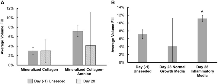
Figure 7.
Mineral formation in mineralized collagen and mineralized collagen–amnion scaffolds in normal growth media. Average mineral fill was quantified by ImageJ processing of micro-CT stacks of mineralized collagen and mineralized collagen–amnion scaffolds. (A) Mineral formation of mineralized collagen scaffolds and mineralized collagen–amnion scaffolds in normal growth media without cells at Day (−1) and with cells at Day 28. No significance was found between all samples. (B) Mineralized collagen–amnion scaffolds in normal growth media and inflammatory media. ^ indicates that the Day 28 inflammatory media group was significantly (P < 0.05) greater than the Day (−1) unseeded group. Data are expressed as mean ± standard deviation (n = 3)

