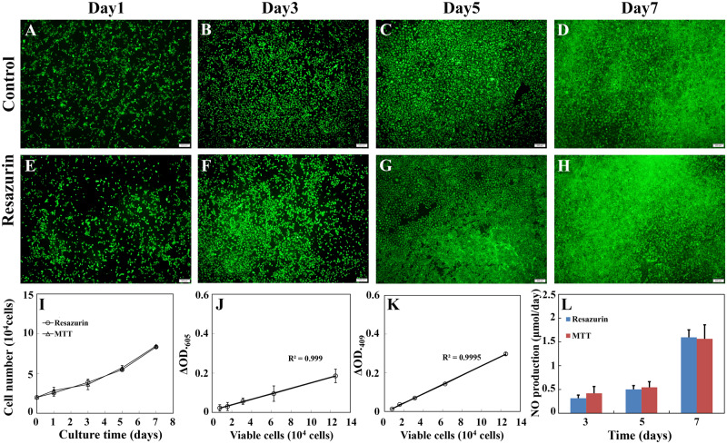
Figure 8.
Effects of repeated viable cell estimation on viability, growth and hemostatic function of RAECs cultured on the sulfated silk fibroin nanofibrous scaffold. (A–H) The representative images of viable cells that were cultured on the sulfated silk fibroin nanofibrous scaffold and determined by live (green)/dead (red) staining. (A–D) Viable cells of the control group. (E–H) Viable cells of the test group, which was monitored by resazurin assay repeatedly. Scale bar: 200 μm. (I) The growth curves of RAECs, which were cultured on the sulfated silk fibroin nanofibrous scaffold. The control group and the test group were monitored by MTT assay and resazurin assay, respectively. The representative standard curves of resazurin assay (J) and MTT assay (K). (L) The productivity of NO of the test group and the control group cultured for 3, 5 and 7 days. Each data was the mean of four scaffolds

