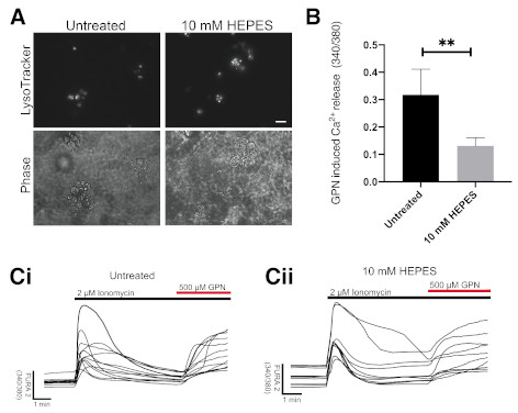
Figure 2. Growth of iPSC-derived neurons in HEPES containing media results in altered lysosomal Ca 2+ and causes lysosomal expansion.

(A) Representative images of iPSC-derived neurons treated for 7 days in media containing 10 mM HEPES. Phase contrast microscopy images show location of neuronal cell bodies. Scale bar = 10 µm, N=3. (B) Following 7-day treatment in HEPES, lysosomal Ca2+ release, triggered by addition of 500 µM GPN, to induce osmotic lysis, after ionomycin to clamp other intracellular Ca2+ stores, was measured in iPSC-derived neurons, N=4 (7–14 cells analysed per repeat). (C) i and ii are Representative traces of Ca2+ release quantified in (B). (*p<0.05, unpaired t-test).
