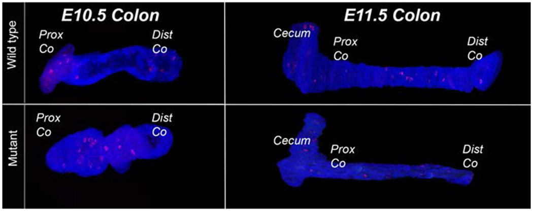
Figure 4: 3D reconstruction of PHH3 staining in epithelial cells of colon.

Three-dimensional reconstruction of colon epithelial tissue from control and Fgfr2IIIb mutant embryos aged E10.5-E11.5. Images reconstructed from 5μm PHH3 stained and images sections of tissue where the mesenchyme in the image was cropped out before reconstruction. There discreet staining found across the length of the epithelium in both the wild type and mutant conditions. Abbreviations: Prox= proximal, Dist= distal, Co = colon
