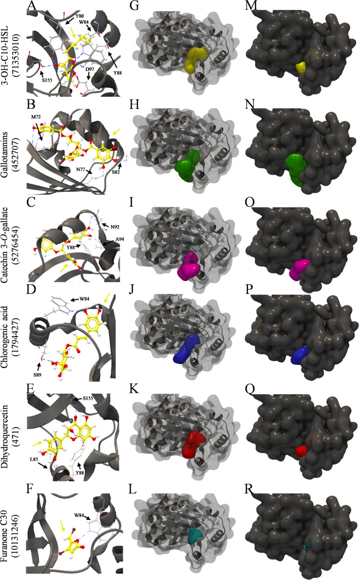
FIGURE 5.
Molecular docking of 3QP8 structure of CviR protein of C. violaceum ATCC 12472 with 3-OH-C10-HSL, gallotannins, catechin 3-O-gallate, chlorogenic acid, dihydroquercetin, and furarone C30. (A–F) Backbone representation of 3QP8 structure with hydrogen bond between the amino acid residues and evaluated compounds, (G–L) surface and backbone representations, and (M–R) surface representation. Gray surface representation, CviR; yellow surface representation, 3-OH-C10-HSL; green surface representation, gallotannins; pink surface representation, catechin 3-O-gallate; blue surface representation, chlorogenic acid; red surface representation, dihydroquercetin; cyan surface representation, furarone C30; gray backbone representation, CviR; black arrow indicates the binding site; yellow arrow, 3-OH-C10-HSL or gallotannins or catechin 3-O-gallate or chlorogenic acid or dihydroquercetin or furarone C30; blue dashed line, hydrogen bond.

