Fig. 1.
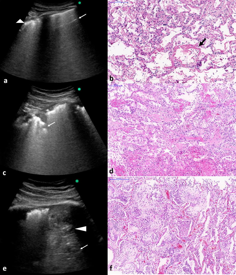
Correlation between lung ultrasound (LUS) postmortem images with histology findings in fatal cases of COVID-19. a, b COVID-19 pneumonia in the early phase with irregular and thickened pleural line (arrowhead) and spared areas with A line (arrow) at LUS examination. The histology shows acute pulmonary injury with hyaline membranes (arrow). c, d intermediary phase with pleural thickening and subpleural consolidations at LUS examination. The histology shows early fibroproliferative changes (in the center) associated with acute diffuse alveolar damage (DAD). e, f LUS examination shows thickened pleural line and consolidation (arrowhead) with air bronchograms (arrow) in the base of left lung. The histology shows fibroproliferative DAD

