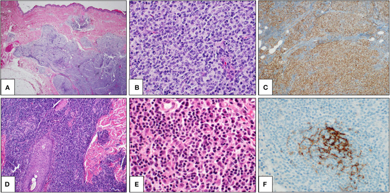
Figure 2.
Primary cutaneous germinal center cell lymphoma: histological findings. Histological examination showing a dense lymphoid infiltrate in dermis and hypodermis, organized in vaguely defined nodules. The lymphoid population infiltrates the hypodermis, entrapping adipocytes (A: H&E, 2×). The lymphoid nodules are constituted by large and irregular germinal centers (B: H&E, 40×) and are positive for bcl6 immunostaining (C: bcl6 immunostain, 10×). Primary cutaneous marginal zone lymphoma: histological findings. A diffuse lymphoid population in the reticular dermis, extending along a hair (D: H&E, 2×). The lymphoid population is heterogeneous, including mature lymphocytes, lympho-plasmacytoid cells, and plasma cells (E: H&E, 40×). CD21 immunostaining highlights a partially destroyed network of follicular dendritic cells (F: CD21 immunostain, 40×).

