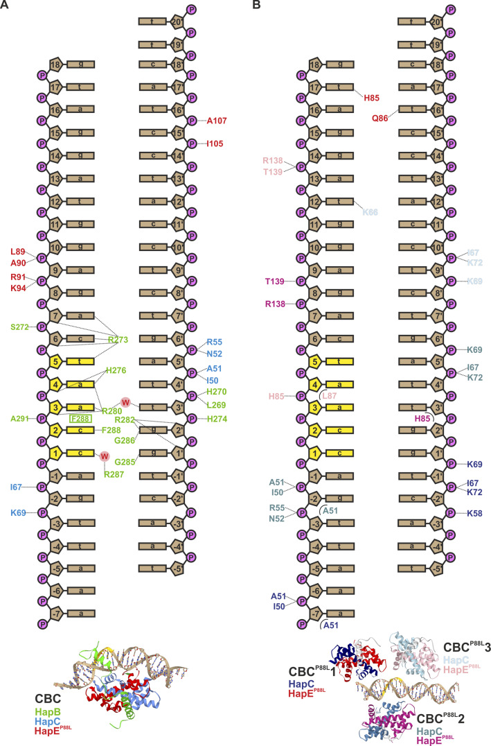
Figure S3. Interactions of wt and HapEP88L-mutant CBC with cycA DNA.
(A, B) Schematic illustration of polar (dotted lines) and apolar (half circles) interactions of wt (A; Huber et al, 2012) and HapEP88L mutant (B) CBCs with the cycA DNA duplex. The CCAAT box is colored yellow. Amino acid labels are colored according to the distinct CBCs (see also the structural scheme below).

