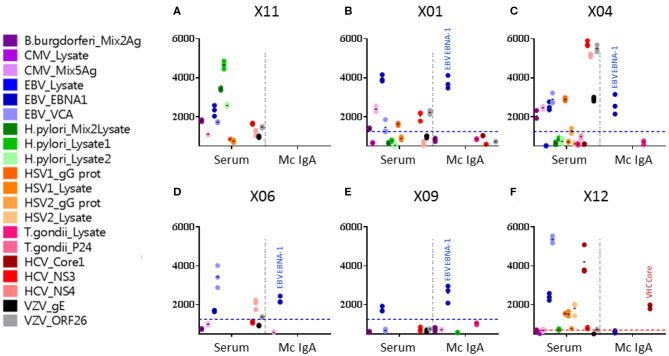
Figure 2.
Viral targets of purified monoclonal IgAs from myeloma patients, as determined by the multiplexed infectious antigen micro-array (MIAA) revealed using a DylightTM 680-labeled goat anti-human IgA Fc antibody. For each patient, serum and purified monoclonal (Mc) IgA were incubated in parallel in the MIAA assay; results shown as fluorescent intensity represent either unseparated IgAs (left) or the patient's monoclonal IgA (right). (A) A patient with a Mc IgA that does not react with any pathogen of the MIAA. (B–E) Four patients with Epstein–Barr virus (EBV)-specific Mc IgAs. (F) One patient with a hepatitis C virus (HCV)-specific Mc IgA. EBV nuclear antigen (EBNA-1) signals are shown in dark blue dots, HCV core signals in red dots, and positive thresholds are shown in dotted lines. (A) For patient X11, the serum contained IgAs that recognized Borrelia burgdorferi, EBV EBNA-1, EBV VCA, Helicobacter pylori lysates 1 and 2, HCV NS3, and varicella zoster virus (VZV) ORF26 protein, whereas the purified Mc IgA did not recognize anything on the MIAA array. (B) For patient X01, the serum contained IgAs that recognized a mix of cytomegalovirus (CMV) antigens, EBV EBNA-1, EBV VCA, herpes simplex virus (HSV-1) gG, HCV NS3, and VZV ORF26; the purified Mc IgA recognized EBV EBNA-1 only. (C) For patient X04, the serum contained IgAs that recognized B. burgdorferi, CMV antigens, EBV EBNA-1, EBV VCA, HSV-1 gG, HCV NS3, HCV NS4, VZV gE, and ORF26, whereas the purified Mc IgA recognized EBV EBNA-1 only. (D) For patient X06, the serum contained IgAs that recognized EBV EBNA-1, EBV VCA, and HCV NS3; the purified Mc IgA recognized EBV EBNA-1 only. (E) For patient X09, both IgAs in serum and the purified Mc IgA recognized the EBV EBNA-1 protein only. (F) For patient X12, the serum contained IgAs that recognized EBV EBNA-1, EBV VCA, HSV-1 gG, HSV-1 lysate, HSV-2 lysate, and HCV core, whereas the purified Mc IgA recognized HCV core only. (B–F) The fluorescence values shown for EBV EBNA-1 or HCV core were obtained after subtraction of the non-specific fluorescent background. Thresholds of specific positivity were defined for each viral pathogen or protein (1,400 for EBV EBNA-1, blue threshold; 500 for HCV core, red threshold) (9, 14, 19). Note that dots may be superimposed; horizontal bars represent the means of results obtained for a pathogen, Ag, or lysate. Experiments were performed in triplicates, repeated at least once.

