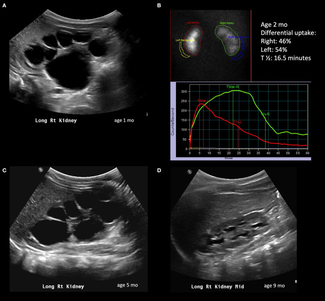
Figure 1.
Radiological history of an infant with resolution of severe hydronephrosis. (A) Postnatal ultrasound confirming prenatally diagnosed hydronephrosis; the image shows SFU grade 4 hydronephrosis with thinned isoechoic parenchyma. (B) Diuretic renogram performed at 2 months of age, showing symmetric uptake; despite the right kidney exhibits a delayed washout curve, it is still considered adequate. (C) Repeat ultrasound at 5 months showing persistent SFU grade 4 hydronephrosis. (D) Follow-up ultrasound at 9 months showing significant spontaneous improvement of hydronephrosis; the child will still however need follow-up imaging to ensure persistent improvement.

