Figure 3.
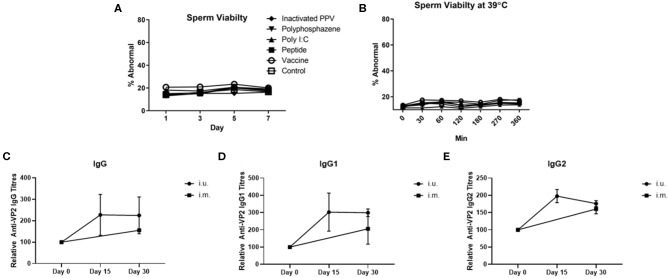
Sperm abnormality assessment of extended semen incubated with intrauterine (i.u.) vaccine components and fetus viability after i.u. immunization and serum antibody titers from animals vaccinated through the i.u. or intramuscular routes (i.m.). Commercially extended semen was incubated alone or in the presence of 1 × 10⁷ TCID50 BEI-inactivated PPV, 400 μg Poly I:C, 800 μg HDP, 400 μg PCEP (i.u. vaccine) at 17°C for 7 days (A) or 39°C (B) with periodic readings for up to 360 min. Acrosome-reacted sperm was bound by peanut agglutinin (PNA) conjugated to Alexa-647 and identified on a FACScalibur. Dead sperm were identified if they were stained with propidium iodide (PI). Experiments were repeated with three separate batches of extended semen. (C–E) Animals were bred with extended semen alone or with i.u. vaccine. Control sows (n = 3) were immunized with ParvoShield vaccine by i.m. route. All sows had previously been vaccinated i.m. with ParvoShield at each breeding cycle when they entered into the farrowing crates (~120 days previously). Serum anti-VP2 IgG (C), IgG1 (D) and IgG2 (E) antibody titres over time were quantified relative to each sow's anti-VP2 titres at day 0 to give relative anti-VP2 titres for i.u.-vaccinated (black circle) and i.m.-vaccinated (black square) sows. Data are presented as means [horizontal bars; (A,B)] and mean with standard deviation in (C–E).

