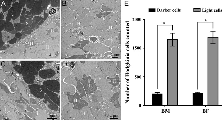
FIG 3.
Localization of primary symbionts and differences in numbers of dark and light “Ca. Hodgkinia” cells in the bacteriomes of Pycna repanda males and females by ultrastructural microscopy. (A) Fragment of the bacteriocytes of males containing “Ca. Sulcia” (S) and “Ca. Hodgkinia” (H). (B) Central part of the bacteriocytes of males containing “Ca. Hodgkinia” cells of different colors. (C) Fragment of the bacteriocytes of females containing “Ca. Sulcia” and “Ca. Hodgkinia.” (D) Central part of the bacteriocytes of females containing “Ca. Hodgkinia” cells of different colors. (E) Numbers of dark and light “Ca. Hodgkinia” cells counted in the bacteriomes of both sexes. We randomly selected multiple sections for statistical analysis, aiming to test the differences in numbers for dark and light “Ca. Hodgkinia” cells in the bacteriomes. Asterisks indicate a statistically significant difference (P < 0.01 by a Mann-Whitney test). BM, bacteriomes of males; BF, bacteriomes of females; N, nucleus.

