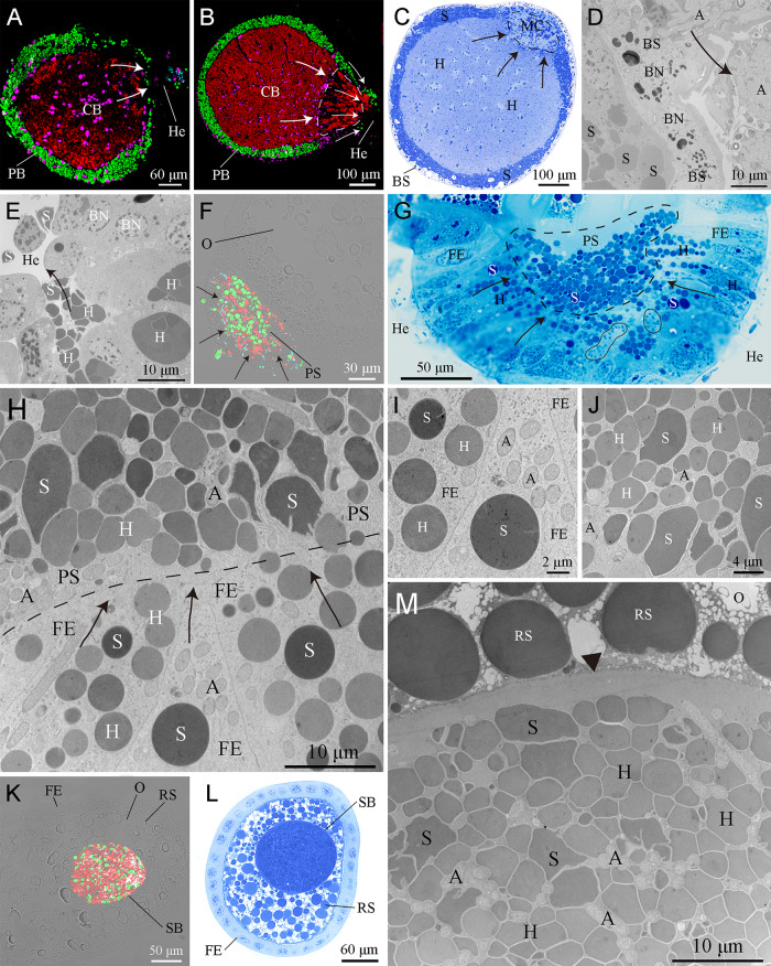
FIG 4.
Transovarial transmission of bacteriome-associated symbionts. (A to E) FISH analysis combined with histological and ultrastructural observations showing Arsenophonus (A), “Ca. Sulcia” (S), and “Ca. Hodgkinia” (H) released from the bacteriocytes into the hemolymph (He). In mature females, Arsenophonus and “Ca. Sulcia” were directly released from the peripheral bacteriocytes (PB) into the hemolymph, but “Ca. Hodgkinia” emigrated from central bacteriocytes (CB) through the multinuclear compartment (MC) (encircled with black/white dotted lines) into the hemolymph. (F to H) Arsenophonus (encircled with a black closed line in panel G), “Ca. Sulcia,” and “Ca. Hodgkinia” toward the posterior pole of the ovarioles, migrating through the cytoplasm of the follicular epithelium (FE) into the perivitelline space (PS) (encircled with a black dotted line in panel G). (I) “Ca. Sulcia” and “Ca. Hodgkinia” remained spherical in the cytoplasm of the follicular epithelium. (J) “Ca. Sulcia” and “Ca. Hodgkinia” seem irregular in the perivitelline space. (K to M) Intermixed Arsenophonus, “Ca. Sulcia,” and “Ca. Hodgkinia” cells forming a characteristic symbiont ball (SB) in the posterior pole of the oocytes (O). Magenta, green, cyan, and red represent the bacteriocyte nucleus (BN), “Ca. Sulcia,” Arsenophonus, and “Ca. Hodgkinia,” respectively. Black and white arrows represent the emigration of the symbionts. The black arrowhead represents the oolemma. BS, bacteriome sheath; RS, reserve substances.

