FIGURE 1.

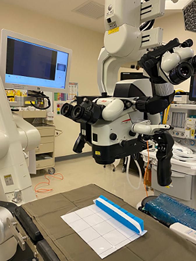
Face shield placed on white background with grid prior to droplet count with surgical microscope [Color figure can be viewed at http://wileyonlinelibrary.com]

Face shield placed on white background with grid prior to droplet count with surgical microscope [Color figure can be viewed at http://wileyonlinelibrary.com]