
Figure 1.
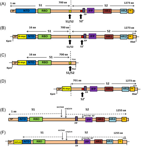
Schematics of spike glycoproteins and recombinant gene constructs. (A) Structure of SARS‐CoV‐2 spike (1273aa) glycoprotein, showing S1 and S2 domains and the cleavage sites S1/S2 and S2′. (B) Structure of pCMV3‐SP‐N‐MYC (Sn). SARS‐CoV‐2 spike (aa16‐aa1273) was cloned into plasmid expression vector at Kpnl and Xbal restriction sites. The N‐terminal 15 amino acids were replaced with signal peptide (SP′) and N‐MYC sequence. (C) Structure of pCMV3‐S1‐N‐MYC (S1‐n). The S1 domain (aa16‐aa700) was cloned into the plasmid expression vector at Kpnl and Xbal restriction sites. The N‐terminal 15 amino acids were replaced with signal peptide (SP′) and N‐MYC sequence. (D) Structure of pCMV3‐S2‐N‐MYC (S2‐n). The S2 domain (aa701‐aa1273) was cloned at Kpnl and Xbal restriction sites. The N‐terminal contains signal peptide (SP′) and N‐MYC sequence. (E) Structure of SARS spike (1255aa) glycoprotein, showing S1 and S2 domains and the cleavage sites S1/S2 and S2′. (F) Structure of p3XFLAG‐CMV‐S (So). SARS spike was cloned into plasmid expression vector as previously described. FP, fusion peptide; HR1, heptad repeat 1; HR2, heptad repeat 2; NTD, nontranslated domain; RBD, receptor‐binding domain; SARS CoV, severe acute respiratory syndrome coronavirus; SP, SARS signal peptide; SP′, signal peptide