
Figure 5.
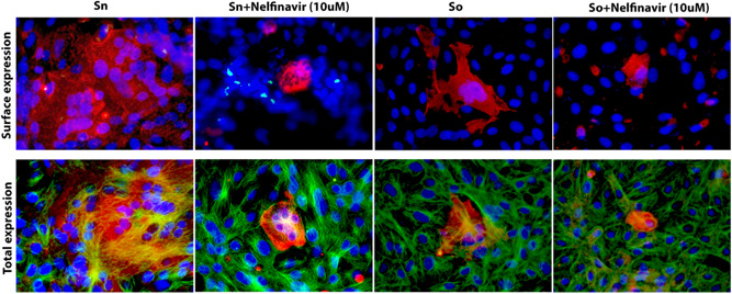
Surface expression of spike glycoproteins. Vero cells were transfected with plasmids expressing either the S‐o or S‐n glycoproteins tagged with the 3XFLAG and N‐MYC epitopes at their amino termini, respectively. S‐n and S‐o expression was detected with mAbs against the epitope tags at 48 hours posttransfection and compared to vehicle containing equivalent amount of DMSO. Nelfinavir was added at the time of transfection at the concentrations indicated. Formalin or Methanol fixed cells were incubated with mouse anti‐N‐MYC (Sn) (1:100) or mouse anti‐FLAG (So) (1:200) antibody and stained with Alexa fluorophore 647 conjugated goat anti‐mouse secondary antibody (1:1000). Cellular tubulin was stained with rabbit anti‐alpha tubulin (Abcam; 1:200) and anti‐rabbit secondary antibody conjugated with Alexa fluorophore 488. DAPI was used to stain nuclei of cells. Fluorescent images were taken at ×40 magnification. DAPI, 4′,6‐diamidino‐2‐phenylindole; DMSO, dimethyl sulfoxide; Sn, S‐new; So, S‐old