
Figure 5.
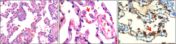
Atypical pneumocytes. A, High‐power view of pneumocyte hyperplasia [haematoxylin and eosin (H&E)]. B, High‐power view showing an enlarged pneumocyte (arrow) with amphophilic granular cytoplasm and a prominent eosinophilic nucleolus (H&E). C, Immunohistochemical staining showing atypical pneumocytes to be positive for pan‐cytokeratin (arrow), confirming their epithelial origin (streptavidin peroxidase). [Colour figure can be viewed at wileyonlinelibrary.com]