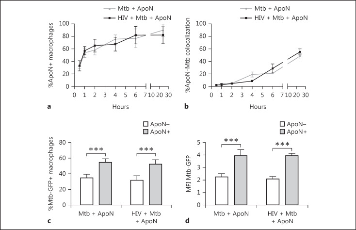
Fig. 2.
Uptake of apoptotic neutrophils by macrophages is time dependent and causes an increase in bacterial phagocytosis. The percentage of macrophages containing apoptotic neutrophils (ApoN+; a) and percentage of colocalization of apoptotic neutrophils to M. tuberculosis phagosomes (b), at the indicated time points, as quantified by microscopy. The macrophages were first infected with HIV followed by M. tuberculosis (Mtb, MOI = 2) infection for 1.5 h, and extracellular bacteria were washed away before stimulation with apoptotic neutrophils for up to 24 h. The graph shows the mean ± SEM from 2 independent experiments. c, d Flow cytometry analysis revealed that phagocytosis of M. tuberculosis (% Mtb-GFP+ macrophages and MFI Mtb-GFP) was increased in infected macrophages containing apoptotic neutrophils (ApoN+) compared to those that were exposed but did not ingest apoptotic neutrophils (ApoN–). Data are the mean ± SEM from 6 independent experiments, where the macrophages were M. tuberculosis infected (MOI = 2) for 2 h followed by apoptotic neutrophil stimulation for 4 h without washing away the bacteria. *** p < 0.001, using repeated-measures ANOVA.

