Fig. 3.
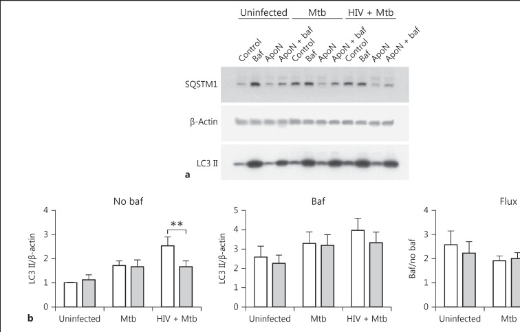
Apoptotic neutrophils do not cause any changes in autophagic flux. a Representative immunoblots showing the autophagy markers LC3B and SQSTM1 (p62), with their β-actin loading controls. The autophagy markers LC3 II (b) and SQSTM1 (c) were quantified from Western blots and normalized to their respective β-actin control and presented as the ratio over uninfected macrophages without apoptotic neutrophils (ApoN). The macrophages were infected with M. tuberculosis (Mtb, MOI = 5) for 1.5 h, washed, and stimulated with apoptotic neutrophils for 22.5 h. “Baf” indicates that the macrophages were pretreated with bafilomycin for 1 h prior to infection, while the graphs named “Flux” show the autophagic flux (i.e., samples with baf/samples without baf). Data are shown as the mean ± SEM with * p < 0.05 and ** p < 0.01 using repeated-measures ANOVA (n = 6). d The percentage of LC3 colocalization to M. tuberculosis was quantified by microscopy after prior HIV infection and 1.5 h of M. tuberculosis infection (MOI = 2) followed by washing and stimulation with apoptotic neutrophils for 4.5 h (6 h in total) or 22.5 h (24 h in total). Data are the mean ± SEM from 4 independent experiments.

