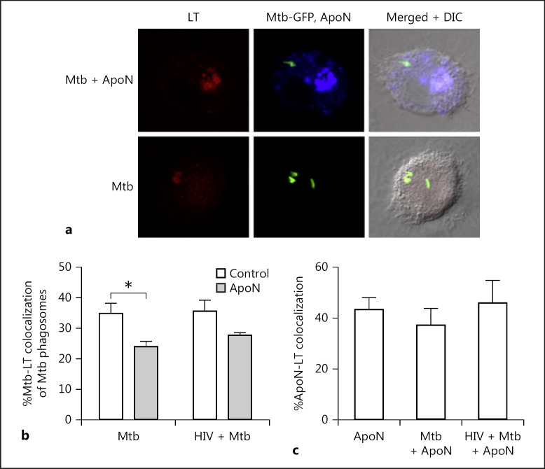
Fig. 4.
Apoptotic neutrophils decrease acidification of M. tuberculosis phagosomes. a Representative micrographs of LysoTracker (LT) colocalization to apoptotic neutrophils (ApoN) or M. tuberculosis (Mtb) phagosomes in infected macrophages. Green, Mtb; red, LT; blue, ApoN. Macrophages were infected with M. tuberculosis for 2 h (MOI = 1), stimulated with apoptotic neutrophils for 4 h, with the addition of the probe LysoTracker (LT) for the last 2 h before fixation and colocalization studies by confocal microscopy. The percentage of colocalization of M. tuberculosis to LT (b) and apoptotic neutrophils to LT (c) for 6 donors, showing the mean ± SEM. * p < 0.05 using repeated-measures ANOVA.

