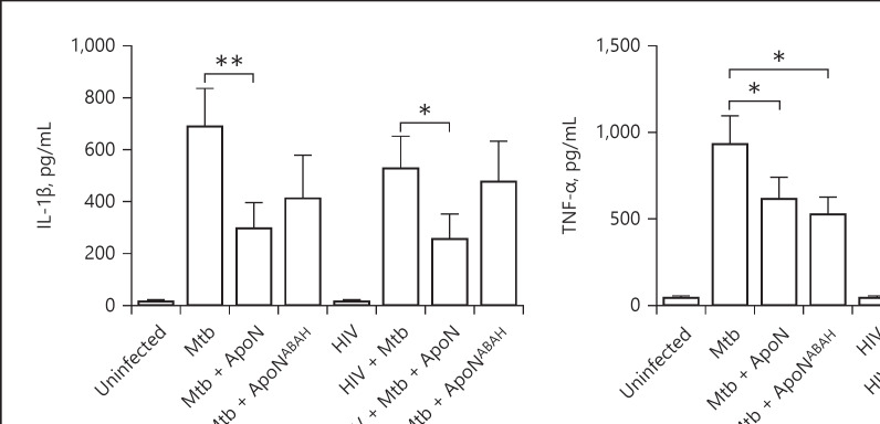
Fig. 8.
Apoptotic neutrophils decrease the proinflammatory response in infected macrophages. IL-1β (a), TNF-α (b), and IL-6 (c) were measured in the supernatants of M. tuberculosis and HIV-coinfected macrophages. Apoptotic neutrophils (ApoN) were treated with 500 μM of ABAH (ApoNABAH) for 1 h prior to washing and added to the infected macrophages. The macrophages were infected with M. tuberculosis (Mtb, MOI = 2) for 1.5 h prior to stimulation with apoptotic neutrophils for 22.5 h. Data are the mean ± SEM from 11 independent experiments. * p < 0.05, ** p < 0.01, using repeated-measures ANOVA with Dunnett's multiple comparison test.

