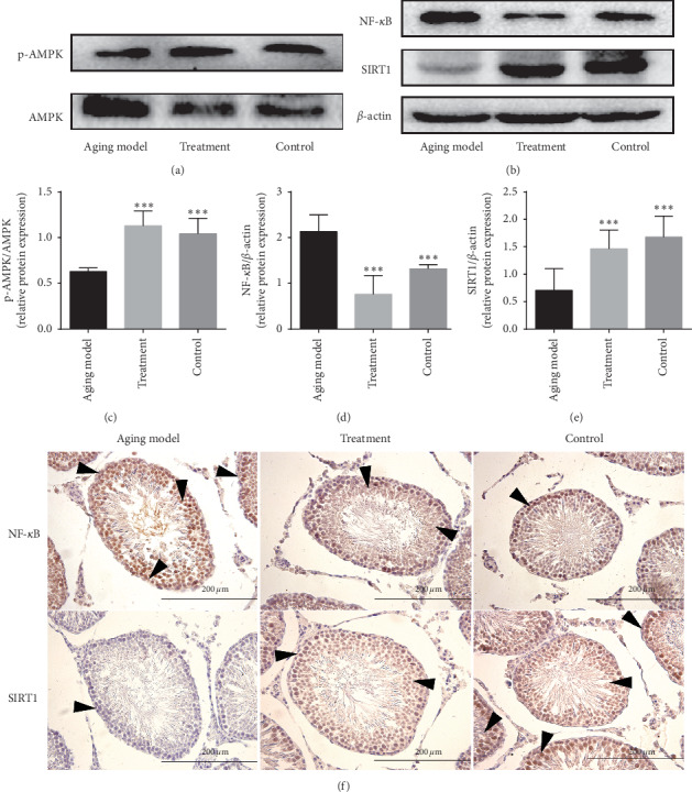
Figure 3.

The effects of LP on anti-inflammation in testicular tissue of aging rats via AMPK/SIRT1/NF-κB. (a) Representative western blot image showing the expression of p-AMPK and AMPK proteins; (b) representative western blot image showing the expression of AMPK, SIRT1, and NF-κB protein; (c) quantification of the ratio of p-AMPK/AMPK; (d) quantification of NF-κB expression; (e) quantification of SIRT1 expression. β-actin was used as an internal loading control. All data are expressed as the mean ± SD, n = 8. ∗P < 0.05, compared with the aging model. (f) Expression of NF-κB and SIRT1 located in the testicular tissue was observed by immumohistochemical staining. Arrows indicate spermatogenic cells stained positive (magnification 400x).
