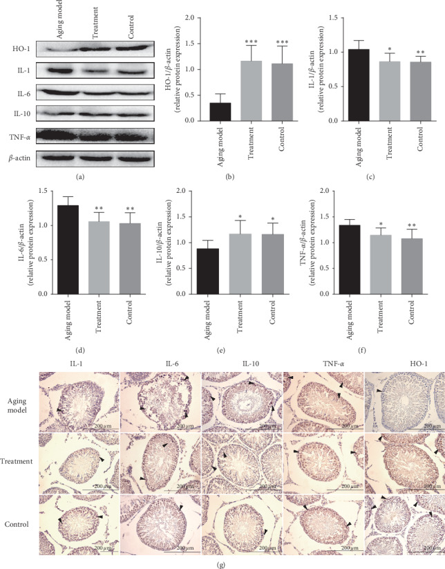
Figure 4.

The effect of LP on anti-inflammation in the testicular tissue was demonstrated by western blot and immunohistochemical staining of testicular tissue of the control group, the aging model, and the treated group. Specific antibodies were used for the detection of IL-1, IL-6, IL-10, TNF-α, and HO-1. (a) Representative western blot image; (d–f) mean densities of HO-1, IL-1, IL-6, IL-10, and TNF-α. All data are expressed as the mean ± SD, n = 8. ∗∗∗P < 0.05, ∗∗P < 0.01, ∗∗∗P < 0.001 compared with the aging model. (g) Representative photographs of immunohistochemical staining. Arrows indicate spermatogenic cells stained positive (magnification 400x).
