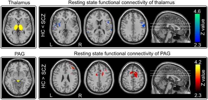
Figure 5.

Resting‐state fMRI functional connectivity. Top panel: Thalamus showed weaker resting‐state functional connectivity with the right S1, right S2, left posterior insula in HC than in SCZ. Bottom panel: PAG had stronger resting‐state functional connectivity with the SMA, dACC, and DLPFC in HC than in SCZ
