
Figure 6.
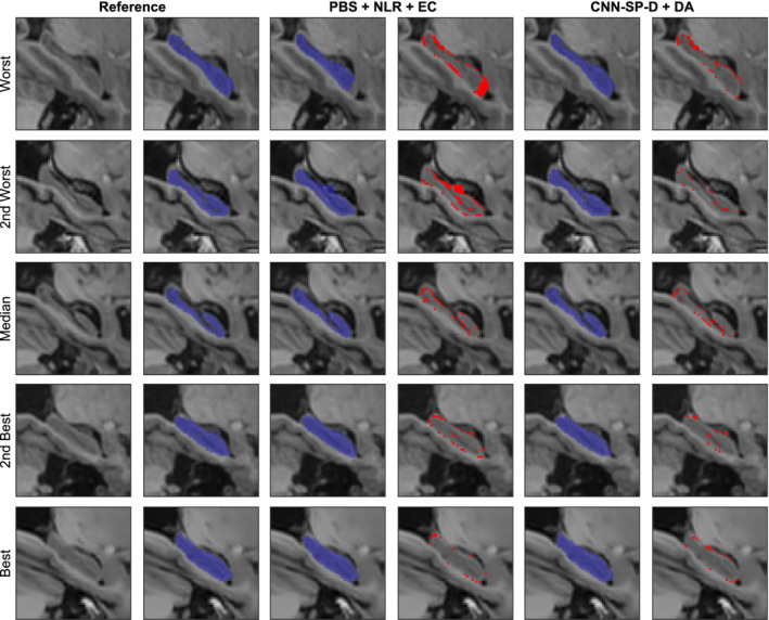
Example right hippocampus segmentations and respective errors using NLR + PB + EC and our best performing method CNN‐SP‐D + DA. The subjects with the worst, second worst, median, second best, and best overlap after applying NLR + PB + EC are shown for comparison. Errors are overlaid in red in columns four and six. EC, error correction; NLR, nonlinear registration; PBS, patch based; CNN‐SP, CNN with spatial priors [Color figure can be viewed at http://wileyonlinelibrary.com]