
Figure 5.
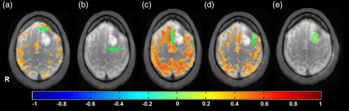
Offline resting‐state analysis in Patient 1 with a glioblastoma using manual seed regions (green) overlaid on T2W magnetic resonance imaging (MRI). Seed locations (a) anterior, (b) posterior, (c) along midline, and (d) lateral to the tumor adjacent to Exner's area display different degrees of connectivity with the sensorimotor and language networks. (e) The seed within the necrotic core of the tumor does not show significant connectivity, as expected