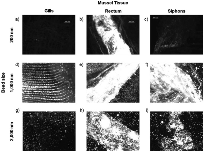
Figure 1.

Exemplar fluorescence images of isolated tissues (gills, rectums, siphons) from quagga mussels dosed with 200 nm (a–c), 1000 nm (d–f), and 2000 nm (g–i) carboxylate‐modified PS beads containing a red dye (excitation/emission 580/605). All images were acquired with the same microscope settings, leading to some images appearing overexposed. The images demonstrate the substantial accumulation of beads of all three sizes in the rectums and of the 1000 and 2000 nm beads in the siphons. Mussels were dosed with 200 and 1000 nm beads at 1 × 10⁻¹² m and 2000 nm beads at 0.01 × 10⁻¹² m for 24 h.
