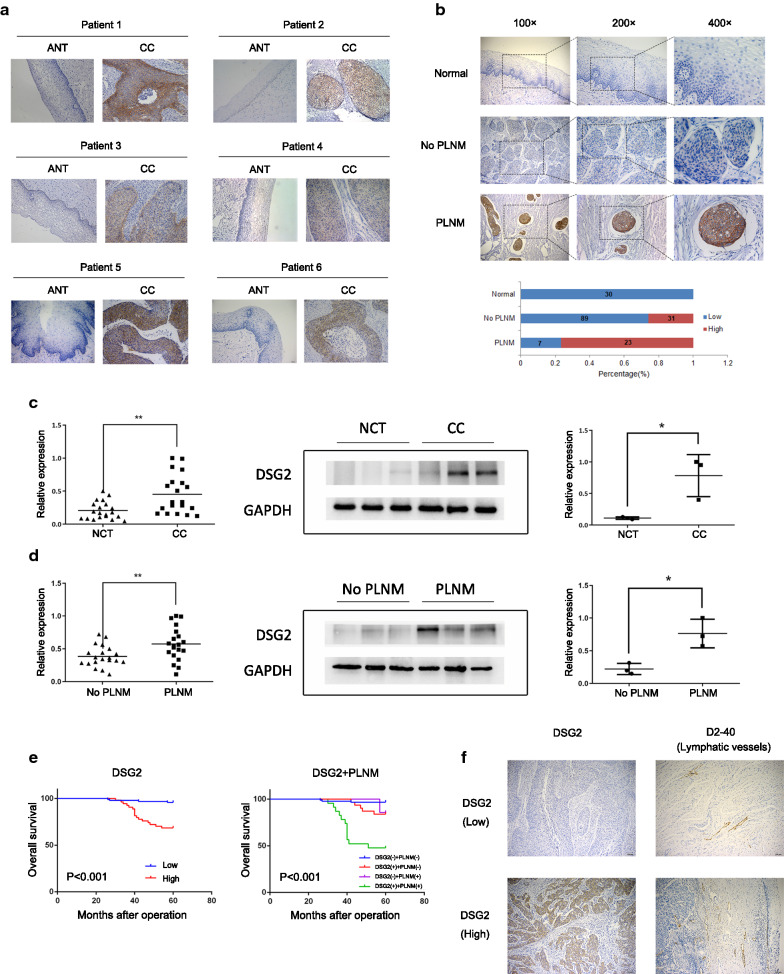
Fig. 3.
The expression of DSG2 in patient samples was determined by IHC, qRT-PCR and western blotting. a IHC was performed on six pairs of matched CC samples and adjacent nontumor cervical tissue (ANT) samples. Original magnifications: ×200. b Representative results of the IHC of DSG2 in normal samples and early-stage CC samples with and without PLNM. Bar graph shows the percentage of high/low expression of DSG2 in normal samples and early-stage cohort samples. c The mRNA expression of DSG2 was determined by qRT-PCR in 20 NCTs and 20 CC samples (left). The protein expression of DSG2 was determined by western blotting in 3 NCTs and 3 CC samples (right). d The mRNA expression of DSG2 was determined by qRT-PCR in 20 CC samples without PLNM and 20 cervical cancer samples with PLNM (left). The protein expression of DSG2 was determined by western blotting in 3 CC samples without PLNM and 3 CC samples with PLNM (right). e Kaplan–Meier curve for DSG2 and PLNM in early-stage cohort CC patients. DSG2 (−): low expression of DSG2; DSG2 (+): high expression of DSG2. PLNM (−): without PLNM; PLNM (+): with PLNM. f Representative results of the IHC of lymphatic vessel density in CC with high/low expression of DSG2. Original magnifications: 100×

