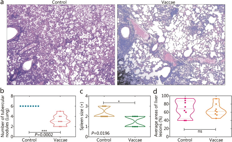
Fig. 4.
Gross pathology and histopatological analysis. The right lobe of lungs collected from the mice in the control group (a, left) and M. vaccae group (a, right) were used to undergo histopathological examination (H&E). The gross pathology of organs was also observed, including the number of the tubercular nodules in the lung b, spleen size average c, and areas of lesions in the liver d. Original magnification times: ×100. All data are presented as means + S.E.M. (n = 8). Differences were considered statistically significant at P < 0.05. *, P < 0.05; ***, P < 0.001; ns. Not significant

