Fig. 4.
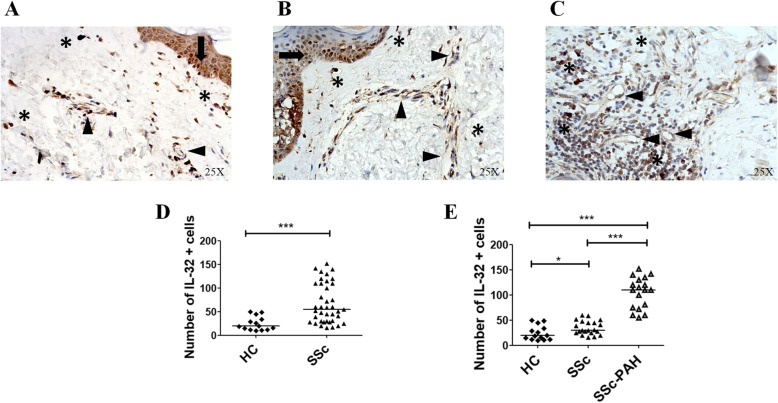
IL-32 expression in the skin of SSc patients. a–c IHC of IL-32 in skin derived from HCs (a), SSc patients (b) and SSc-PAH patients (c). IL-32 was expressed in keratinocytes (arrow), vascular cells (arrowhead) and fibroblasts (*). Original magnification × 25. Negative controls were obtained by omitting the primary antibody. d Quantification of the number of IL-32+ cells in HCs and SSc patients. The number of IL-32+ cells are significantly increased in SSc patients when compared to SSc (***p = 0.0001). e Quantification of the number of IL-32+ cells in 14 HCs, 21 SSc patients without PAH and 18 SSc patients with PAH. In SSc-PAH patients, the number of IL-32+ cells is significantly increased when compared to SSc patients without PAH. Any dot plot is representative of the median cells count per 2 section for each patient (*p = 0.02; ***p < 0.0001)

