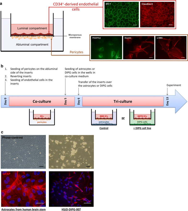
Fig. 1.
Modelling of the human blood- brain tumor barrier in pediatric DIPG. a Shematic representation of the syngeneic human in vitro BBB model where endothelial cells (ECs) were differentiated from human cord blood CD34+ hematopoietic stem cells and cultured on the luminal side of the insert. ECs were co-cultured with human brain pericytes, seeded on the abluminal side of the insert. Cells were visualized by performing immunostainings of platelet-derived growth factor receptor-β (PDGFR-β), desmin and α-smooth muscle actin (α-SMA) for pericytes, and tight junction proteins zonula occludens-1 (ZO-1) and claudin-5 for ECs. Scale bar = 25 µm. b Synopsis of culture, starting on day 0 with the seeding of ECs and pericytes on the luminal side and the abluminal side of the insert, respectively. After 6 days of co-culture, ECs and pericytes were incubated with either the astrocytes, to model the healthy BBB (control), or the DIPG cells, to model the blood–brain tumor barrier (+ DIPG cell line), for a duration of 7 days. c Representative images of glial fibrillary acid protein (GFAP) immunostainings on human astrocytes and DIPG-007 cells. For phase-contrast pictures, scale bar = 50 µm. For immunofluorescence pictures, scale bar = 25 µm

