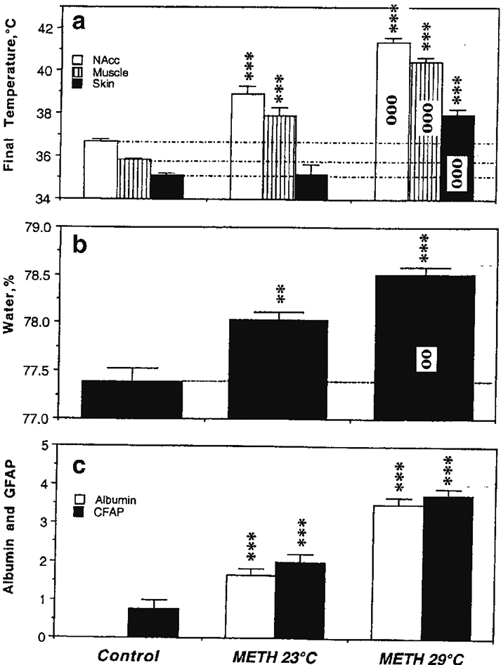
Figure 2.

Mean (±SEM) values of temperature (a), brain tissue water content (b) and immunoreactivity for albumin and GFAP (c) in rats exposed to METH in standard (23°C) and warm (29°C) environmental temperatures. Asterisks show values significantly different from the control (intact rats that received saline injections), and circles indicate significant differences between 23 and 29°C. Original data were published in Kiyatkin et al., 2007.
