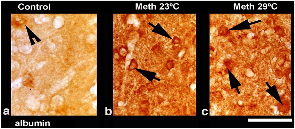
Figure 4.

Leakage of serum albumin in the cerebral cortex in rats exposed to METH at 23° C (b) and 29°C (c) as compared to the saline-treated control (a). In METH-treated rats, albumin positive cells are seen within the neuropil (arrows) largely in the neurons (b and c). Distorted neurons with perineuronal edema were more frequent in rats that received METH at 29°C compared to 23°C. Data were modified after Sharma and Kiyatkin, 2010.
