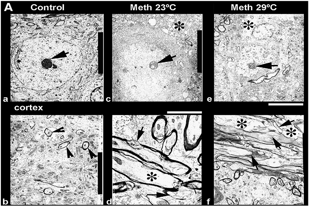
Figure 7.

Ultrastructural changes in the cortex (a, c, e) and thalamus (b, d, f) after METH treatment at 23°C (c, d) and 29°C (e, f) as compared to the control (a, b). Degeneration of the neuronal nucleus and eccentric nucleolus (arrow) is clearly seen in METH-treated rats, and the magnitude and intensity of cellular and membrane degeneration was higher at 29°C as compared to 23°C (a, c, e). In the thalamus, myelin vesiculation (d, f, arrows), expansion of neuropil (*) and membrane disruption were quite common in the METH-treated group. The intensity of these changes was more aggravated by METH treatment at 29°C (f) as compared to identical treatment at 23°C (d). Data are modified from Sharma and Kiyatkin, 2010.
