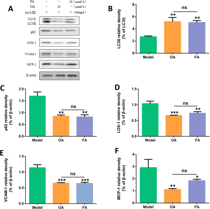
Fig. 10.
Comparison of protective effects of FA and OA on injured HUVECs. a Protein expressions of LC3II, p62, LOX-1, VCAM1, MCP-1, β-actin in HUVECs. The cells were treated with 40 mg/L ox-LDL (model). In addition, cells were pretreated with OA or FA for 2 h followed by 40 mg/L ox-LDL exposure for another 24 h. b, c, d, e, f Bar charts show the mean intensity of every protein quantified and normalized versus β-actin expression. Values are submitted as mean ± SD based on seperate experiments in triplicate.(*) P < 0.05, (**) P < 0.01 and (***) P < 0.001 versus model

