Fig. 3.
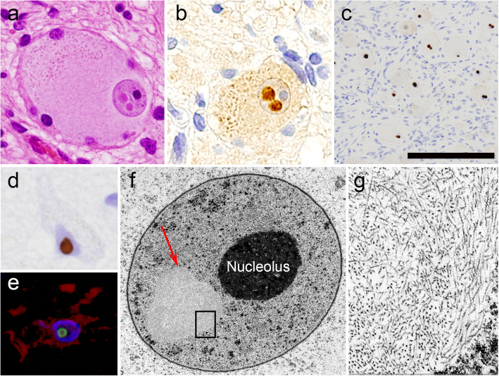
Intranuclear inclusions in extra-skeletal muscle organs. a-c Sympathetic ganglia. Two eosinophilic neuronal intranuclear inclusions with surrounding halos are evident on HE staining (a). The inclusions are positive for ubiquitin (b). Numerous inclusions are revealed by p62-immunohistochemistry (c). d A p62-positive neuronal intranuclear inclusion in the transentorhinal cortex. e A p62-positive intranuclear inclusion (green) in a GFAP-positive astrocyte (red) in the temporal cortex. Double-labeling immunofluorescence. f, g Ultrastructure of a sympathetic ganglion neuron containing an intranuclear inclusion (f, arrow). The inclusion is composed of fine filamentous structures without limiting membranes. Scale bar: a, b = 40 μm; c = 150 μm; d, e = 25 μm; f = 5 μm; g = 750 nm

