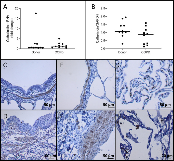
Fig. 5.
Cathelicidin expression No significant difference in cathelicidin mRNA levels were observed between COPD explant tissue and tissue from unused donor lungs (p = 0.35). b. Cathelicidin protein expression was not different in COPD explant tissue compared to tissue from unused donor lungs (p = 0.09). Negative cathelicidin staining in bronchial epithelium with positive smooth muscle layer of an unused donor lung (c) and COPD explant lung (d). Negative endothelial staining with positive smooth muscle layer in an unused donor lung (e) and COPD explant lung (f). Positive immune cell staining in an unused donor lung (g) and COPD explant lung (h). Representative images are shown. N = 10 Horizontal line represents the median value

