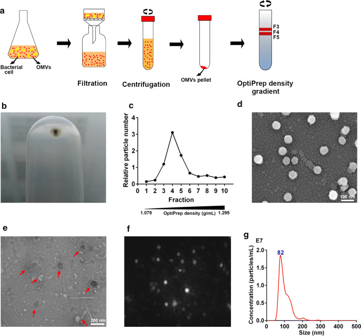
Fig. 1.
Preparation and visualization of OMVs derived from avian pathogenic Escherichia coli. a Isolation and purification protocols of bacterial OMVs. b Native OMVs were isolated and pelleted by ultracentrifugation. c OMVs were purified by Optiprep density gradient ultracentrifugation. The particle numbers of the resulting fractions (1–10) were detected by nanoparticle tracking analysis (NTA). Purified MOMVS were visualized using scanning electron microscope (d) and transmission electron microscopy (e) after negative staining. f Representative frame was captured from the MOMVs NanoSight videos. g Size distribution and concentration of these vesicles was determined by NTA

