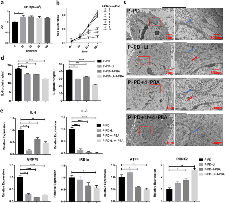
Fig. 3.
LIPUS may reduce the expression of inflammatory factors in P-PDLSCs by inhibiting ERS. a, b CCK-8 assay optimized the intensity of LIPUS (a) and the dose of 4-PBA (b) (n = 5). c TEM images observed the morphology of ER. The red box is the area of interest, the blue arrow indicates the location of ER, and the red arrow refers to the autophagosome. d ELISA was used to detect the secretion of IL-6 and IL-8 inflammatory factors (n = 3). e LIPUS and 4-PBA modulated the levels of IL-6, IL-8, GRP78, IRE1α, ATF4, and Runx2 by qRT-PCR analysis (n = 3). Gene expression was normalized to GAPDH. All data are presented as means ± SD. The representative results were from three independent experiments (ns, no significant; *p < 0 .05; **p < 0 .01; ***p < 0 .001; ****p < 0 .0001). P-PD, P-PDLSCs; H-PD, H-PDLSCs; LI, LIPUS; H, hour

