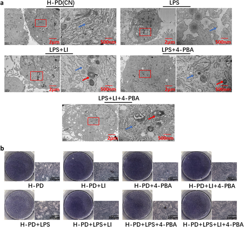
Fig. 6.
LPS caused ERS and reduced the osteogenic capacity of H-PD. a Protective effect of LIPUS on the ultrastructural changes in LPS-induced H-PDLSCs by TEM: LPS (10 μg/ml), 4-PBA (5 μM), and LIPUS (90 mW/cm2) were used. Well-arranged rough endoplasmic reticula (RERs) with abundant attached ribosomes were observed in the control group. Rapid proliferation of RERs, some swelled cisternae, and degranulation were shown in cells treated with LPS. Blue arrows indicated ultrastructural changes of ER in H-PDLSCs. Red arrows indicated the autophagosome. b ALP staining was used to detect osteogenic differentiation for different groups. The representative results were from three independent experiments. P-PD, P-PDLSCs; H-PD, H-PDLSCs; LI, LIPUS

