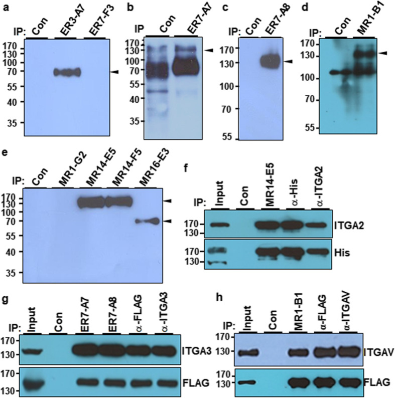
Fig. 2.

Identification of cell-surface molecules recognized by selected MAbs. a–e A549 cell lysates were immunoprecipitated with the indicated MAbs after cell surface biotinylation. After bead-bound proteins were eluted, the proteins were detected with SA-HRP in Western blotting. Preclearing was done with protein G agarose beads and used as a negative control. f Overexpression and immunoprecipitation of integrin α2 (ITGA2) with MR14-E5, anti-His, or anti-ITGA2 antibodies. HEK293T cells were transfected with pcDNA3.1+ (control vector) or pCMV-His-ITGA2 expression plasmids, and the cell lysates were immunoprecipitated with MR14-E5, anti-His, or anti-ITGA2 antibodies. The immunoprecipitates were analyzed by Western blotting with anti-ITGA2 or anti-His antibodies. g Overexpression and immunoprecipitation of integrin α3 (ITGA3) with ER7-A7, ET7-A8, anti-His, or anti-ITGA3 antibodies. HEK293T cells were transfected with pcDNA3.1+ or pCMV-FLAG-ITGA3 expression plasmids, and the cell lysates were immunoprecipitated with ER7-A7, ER7-A8, anti-FLAG, or anti-ITGA3 antibodies. The immunoprecipitates were analyzed by Western blotting with anti-ITGA3 or anti-FLAG antibodies. h Overexpression and immunoprecipitation of integrin αV (ITGAV) with MR1-B1, anti-FLAG, or anti-ITGAV antibodies. HEK293T cells were transfected with pcDNA3.1+ or pCMV-FLAG-ITGAV expression plasmids, and the cell lysates were immunoprecipitated with MR1-B1, anti-FLAG, or anti-ITGAV antibodies. The immunoprecipitates were analyzed by Western blotting with anti-ITGAV or anti-FLAG antibodies. Preclearing was done with protein G agarose beads and used as a negative control
