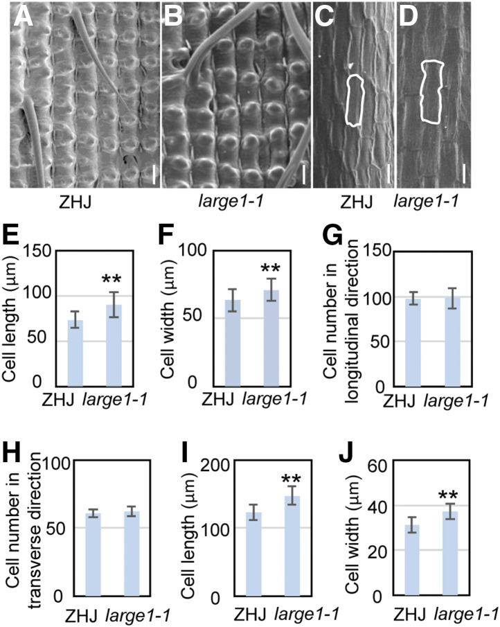
Figure 2.
large1 Forms Large Grains Due to Increased Cell Expansion in the Spikelet Hull.
(A) and (B) Scanning electron microscopy analysis of the outer surface of ZHJ (A) and large1-1 (B) lemmas.
(C) and (D) Scanning electron microscopy analysis of the inner surface of ZHJ (C) and large1-1 (D) lemmas.
(E) and (F) Average length (E) and width (F) of outer epidermal cells in ZHJ and large1-1 lemmas.
(G) Outer epidermal cell number in the longitudinal direction in ZHJ and large1-1 lemmas.
(H) Outer epidermal cell number in the transverse direction in ZHJ and large1-1 lemmas.
(I) and (J) Average length (I) and width (J) of inner epidermal cells in the longitudinal direction in ZHJ and large1-1 lemmas.
Values ( [E] to [J]) are given as the means ± sd (n ≥ 50). **, P < 0.01 compared with the wild type by Student’s t test.
Bar in (A) to (D) = 50 µm.

