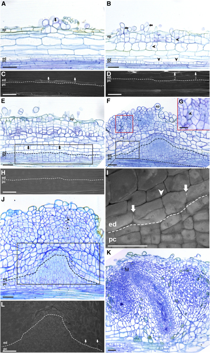
Figure 1.
Parasponia Root Nodule Developmental Stages.
(A) Nodule formation starts with cell divisions in the epidermis (arrow).
(B) Cell divisions are induced in the outer cortical cell layers and pericycle (arrowheads), and divisions continue in the epidermis (arrows).
(C) and (D) UV light images of the endodermis regions of the sections shown in (A) and (B), respectively, to visualize the fluorescence of Casparian strips. At the site of nodule primordium formation, the Casparian strips of endodermis cells are visible (arrows).
(E) Cell divisions occur in all cortical cell layers, pericycle, and the endodermis (arrows).
(F) and (G) Cortex-derived cells contain rhizobium-induced infection threads (see arrowhead in [G]) and the red boxed region in [F]). Pericycle-derived cells form a dome-shaped structure flanked by root endodermis-derived cells.
(H) and (I) UV light images of the root endodermis regions indicated by the black boxes in the sections shown in (E) and (F), respectively.
(H) Mitotically activated root endodermis cells have lost their Casparian strips.
(I) Casparian strips reformed in the peripheral region (arrows; see Supplemental Figure 4B for the right-side region) but are still absent in the central region of root endodermis-derived cells (Supplemental Figure 4C). The root endodermis-derived cells have divided, especially the cells between the two arrows in (I). The Casparian strips are not present at the conjunction site between these two dividing endodermal cells (arrowhead). This implies that these Casparian strips (arrows) are newly formed.
(J) Cell divisions continue in the outer cortical cells. The pericycle-derived dome-shaped structure starts to elongate and forms the vascular tissues. At the apex of the pericycle-derived vasculature, endodermis-derived cells have a thinner cell wall and appear to remain mitotically active.
(K) The outer cortex-derived cells become part of the mature nodule. A group of dividing cells at the apex of the nodule vasculature appears to form a nodule meristem (M), supporting the growth of the nodule vasculature and adding cells to the infected tissue.
(L) UV light image of the root endodermis region of the section shown in (J). Root endodermis-derived cells flanking the pericycle cells have formed Casparian strips (arrows). This is the newly formed endodermis that surrounds the nodule vasculature. Endodermis-derived cells remain undifferentiated at the apex of the pericycle-derived vasculature and appear to maintain mitotic activity. Dotted lines mark the border between endodermis- and pericycle-derived cells. ep, Epidermis; ed, endodermis; pc, pericycle. Scale bars, 50 µm ([A] to [F] and [H] to [L]), 25 µm (G).

