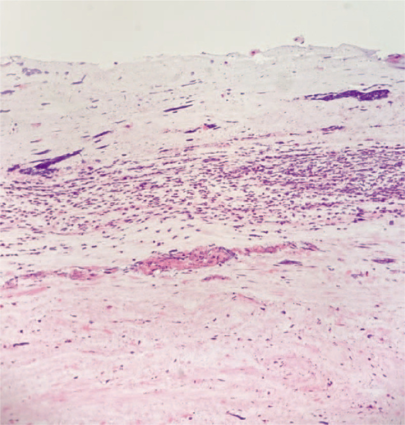
FIGURE 2.

Demonstrated is a microscopic image (20× magnification) of a patient's ETT contents with a trilaminar appearance with a superficial layer of mucin, an underlying layer of degenerated inflammatory cells, and a deep collagenous layer with spindle cells suggestive for stroma. ETT indicates endotracheal tube.
