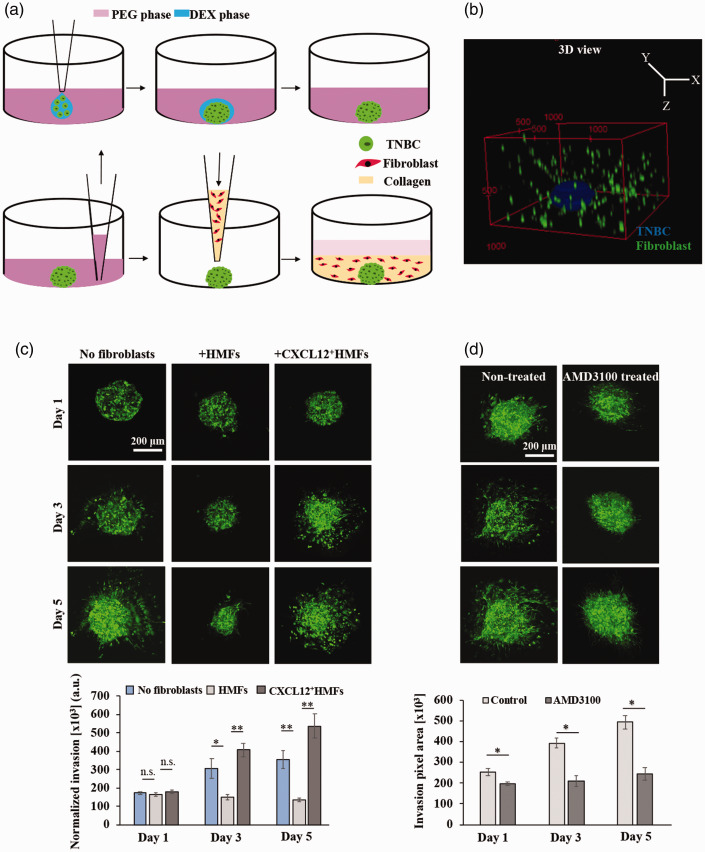
Figure 4.
(a) Schematics of formation of organotypic tumor model in two convenient steps: first forming spheroids using the ATPS technology and then embedding it in a collagen hydrogel containing dispersed fibroblasts. (b) 3D reconstructed confocal image of the tumor model. Note that collagen is not shown. (c) Confocal z-projected images of TNBC cells and quantified matrix invasion of TNBC cells in the tumor models with and without fibroblasts. (d) Confocal images of TNBC cells with and without AMD3100 treatment and the quantified invasion results. *p < 0.05, **p < 0.001. This figure is reproduced with permission from Biomaterials, 2020; 238:119853. (A color version of this figure is available in the online journal.)

