Figure 2.
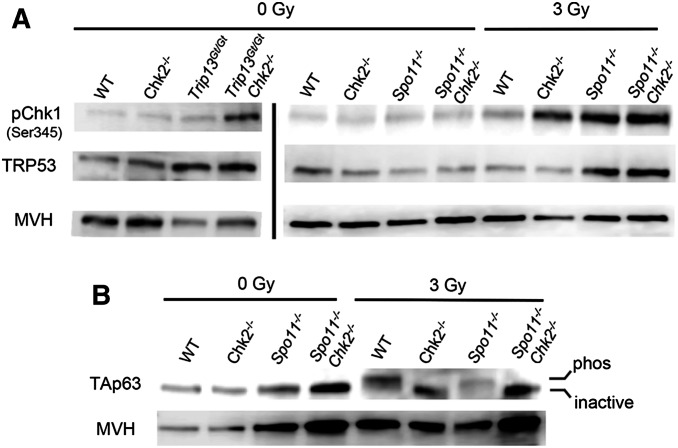
Increased CHK1 activation and p53 stabilization, but not TAp63 activation, in CHK2-deficient oocytes. (A) CHK1 phosphorylation in oocytes is stimulated by induced or meiotic DSBs. Shown are Western blots probed with indicated antibodies. Each lane contains total protein extracted from four ovaries (postnatal day 3–5) that were either exposed or not to 3 Gy of ionizing radiation (IR). Ovaries were harvested for protein extraction 3 hr post-IR. The blots on the left, separated by a vertical bar from those on the right, were from a different blot and different protein samples and mice. The same two blots (left and right) were stripped and reprobed sequentially with the three antibodies. A biological replicate is shown in Figure S1. Note that the decreased MVH levels in Trip13Gt/Gt ovaries is due to reduction in oocytes. (B) Activation of the TAp63 isoform is dependent on DNA damage and CHK2 signaling, not asynapsis. Shown is a Western blot probed sequentially for TAp63 and the germ cell marker MVH. Each lane contains protein extracted from ovaries as described in A. An upward shift in the band indicates the presence of the active (phosphorylated) vs. inactive TAp63. A biological replicate is shown in Figure S1.

