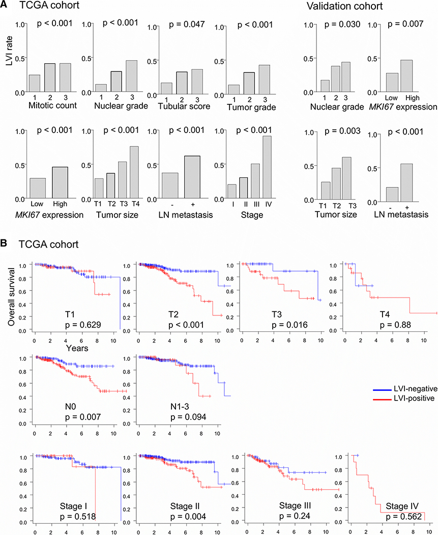
Figure 2.
Lymphovascular invasion (LVI) and clinical and pathological features. a LVI incidence rates (fractions) in the TCGA-BRCA and validation cohorts are plotted for different features. P values were determined with Fisher’s exact test. Median value was used to classify tumors into two groups by their MKI67 gene expression. LN, lymph node. b Kaplan-Meier survival plots along with log-rank test p values are shown for overall survival among LVI-positive and -negative cases of the TCGA cohort sub-grouped by TNM T and N status and pathologic stage.

