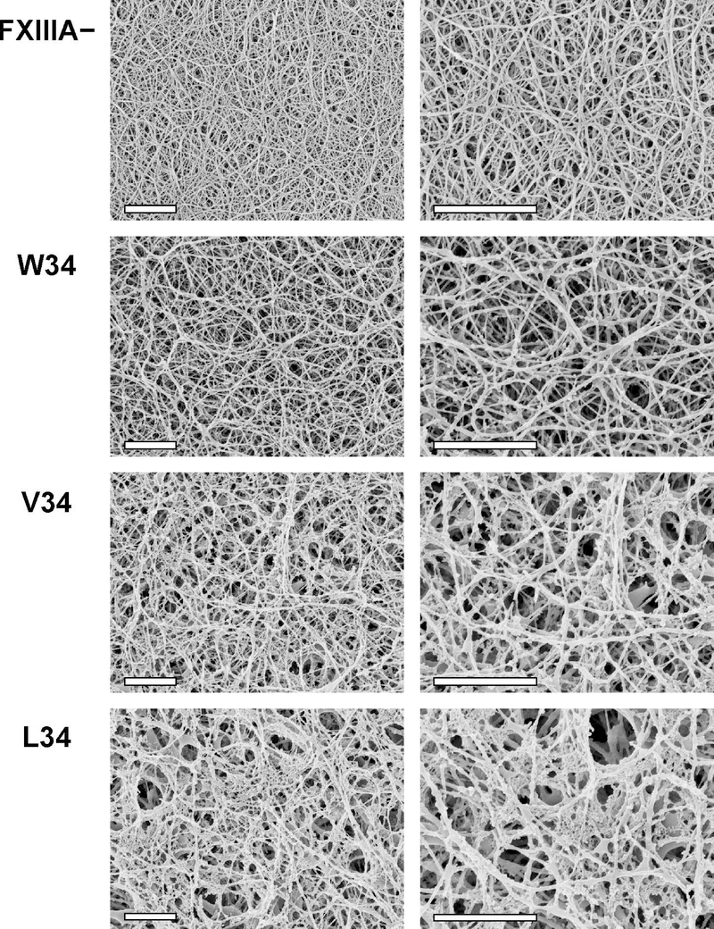
Fig. 3. Scanning electron microscopy of fibrin clots in the presence of FXIIIA AP variants.
100 nM FXIIIA AP variants were combined with plasma from FXIIIA-deficient mice (final dilution of plasma was 1:4). Control samples were made without FXIIIA (FXIIIA −). Clotting was initiated by addition of 2.1 NIH units/ml bovine thrombin and 13.5 mM CaCl2. Clots were formed for 2 h at 37 °C and prepared for SEM as described in Materials and Methods. For each FXIIIA AP variant, two clots were studied, with essentially the same results. Each set of clots (FXIII −, W34, V34, and L34) was formed using plasma obtained from the same mouse. Shown are representative SEM photographs at two magnifications: 10,000x (left) and 20,000x (right), scale bars are 2 μm. Note that during the SEM sample preparation process, the fixative agent glutaraldehyde was not included to avoid artificial protein crosslinking. As a result, the FXIIIA− clots were significantly flattened during the dehydration process. In response, the resultant FXIIIA− fibrin fibers on the SEM images appeared denser packed.

